The treatment of chronic myeloid leukemia (CML) has advanced dramatically with the advent of tyrosine kinase inhibitors (TKIs). However, achieving a true functional cure remains far from straightforward. One of the major obstacles is the persistence of leukemic stem cells, which can remain in the body even after treatment.
How can researchers identify these rare cell populations, clarify their properties, and ultimately find ways to eliminate them from the body? The research team led by Dr. Tomoiku Takaku and Dr. Kohjin Suzuki at the Department of Hematology, Juntendo University Graduate School of Medicine — now affiliated with the Department of Hematology at Saitama Medical University Hospital — has spent many years working on this challenge. Yet the team continued to encounter a barrier that conventional flow cytometry could not overcome.
This article retraces their journey of how they encountered Ghost Cytometry® (GC) technology and began moving beyond the limitations of conventional analysis that depends on cell surface markers such as CD markers.
Index
- Challenge: Confronting the Limitations of CD Markers
- Solution: Morphology as a New “Marker”
- Results: Quantifying Morphological Abnormalities – Moving from Intuition to Impact
- Future Outlook: GC is Transforming Drug Discovery Research
- Taking the Next Step
Challenge: Confronting the Limitations of CD Markers
Flow cytometry is an indispensable tool in hematology research, particularly for identifying leukemic stem cells. However, conventional analytical technologies have an inherent limitation: cell classification relies heavily on fluorescent staining using antibodies against CD markers.
“In basic research using mouse models, genetic manipulation makes it possible to label and validate specific cells. But when analyzing human patient samples with clinical application in mind, that approach is not feasible. To capture the true heterogeneity of human leukemic stem cells, analytical methods that rely only on existing markers had clear limitations,” Dr. Takaku recalled.
The inability to sufficiently narrow down the target cells became a bottleneck, limiting improvements in analytical precision and, ultimately, slowing progress in leukemic stem cell research.
From the perspective of a principal investigator, another challenge had also become apparent. If laboratories around the world are focusing on similar markers, there is a limit to the insights that can be gained through those approaches alone.
“If we simply continued pursuing the same targets using conventional methods, it would have been difficult to differentiate our work from other research groups or generate a real breakthrough. To establish the originality of our research, we needed an entirely new technological approach,” said Dr. Takaku.
Solution: Morphology as a New “Marker”
ThinkCyte’s GC technology offered the potential to add a new perspective to leukemic stem cell research. When Dr. Takaku encountered the technology at the Japan Cytometry Society meeting in 2019, he immediately sensed its innovative potential.
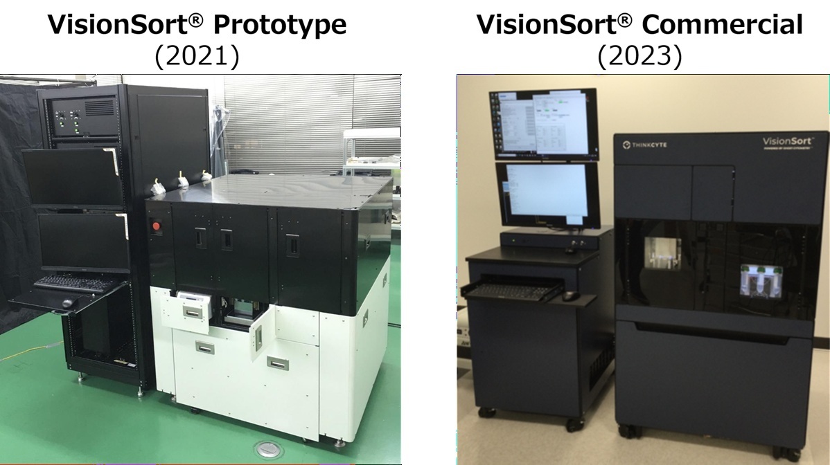
Editor’s note: The research findings discussed in this interview were obtained using a prototype instrument equipped with GC technology. This technology has since been commercialized as the next-generation cell sorter VisionSort®.
The key feature of GC is its ability to analyze and classify cells at high speed and without labels, using AI to interpret cellular morphological information captured through proprietary technology. By overcoming the low-throughput limitations of conventional imaging flow cytometry, GC can rapidly process large numbers of cells, detect structural features that are difficult for the human eye to recognize, and sort cell populations that possess those features.
“What we were looking for was a new indicator beyond the framework of conventional antibody staining. We thought GC might allow us to use AI to objectively express, in numerical terms such as SVM scores or ROC-AUC, the hard-to-verbalize intuition that experienced physicians and technologists develop under the microscope — the sense that ‘something is different about this cell,’” said Dr. Takaku.
“The final deciding factor was not only the expectation that GC’s uniqueness could help differentiate our research from others, but also the promising data we obtained from preliminary experiments using clinical CML samples. Those early results showed high discriminative performance for CML cells and became the driving force behind the full-scale launch of a collaborative research project using a GC-equipped instrument,” he continued.
In adopting the technology, Juntendo University’s extensive clinical expertise and ThinkCyte’s advanced engineering capabilities created a strong synergy.
“We had a deep understanding of clinical needs and medical knowledge, but we did not have sufficient technical expertise in cutting-edge cell analysis instruments. ThinkCyte, on the other hand, was seeking insight into how its proprietary technology could be applied to clinical research. This mutually complementary relationship became one of the strengths that made the collaboration successful,” said Dr. Takaku.
By combining the clinical perspective of hematologists with an engineering approach based on AI-driven morphological analysis, the collaboration established a robust platform for identifying target cell populations through a new approach.
Results: Quantifying Morphological Abnormalities – Moving from Intuition to Impact
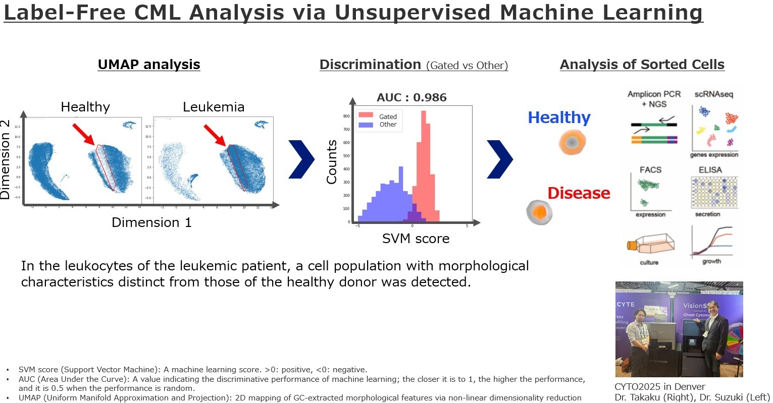
The moment when the value of GC technology became most tangible was when the existence of an unknown cell population identified by GC was convincingly supported by electron microscopy. GC extracted a morphologically distinct cell population that previous approaches had overlooked. When the team observed those cells by electron microscopy, they confirmed a clear biological feature: mitochondrial fragmentation.
“When the classification results from GC were supported by established technologies such as electron microscopy, we truly felt how compelling this technology could be,” said Dr. Takaku.
The introduction of GC produced promising outcomes in both scientific discovery and research strategy.
- Quantifying “clinical intuition”: Differences in cellular morphology that had previously been recognized only intuitively can now be defined objectively and quantitatively through the use of AI. “For example, in diseases such as myelodysplastic syndromes (MDS), where morphological abnormalities are decisive for diagnosis, GC may make it possible to quantify morphological differences,” said Dr. Takaku. “Morphological discrimination that previously had to rely on subjective judgment can now be processed statistically at the level of thousands to tens of thousands of cells. By applying this approach, we are beginning to see the possibility of using it as an objective indicator for early diagnosis or prediction of treatment response. It has also expanded our thinking toward applying the approach to a range of diseases beyond CML,” said Dr. Suzuki.
- A role as a novel marker: GC can visualize additional heterogeneity within cell populations that would previously have been regarded as homogeneous based on specific CD markers. Rather than being simply an analytical instrument, GC has the potential to be positioned as a new kind of marker that follows cell surface markers, said Dr. Suzuki.
- Improving research visibility: Incorporating cutting-edge AI × cytometry technology into the research also proved effective for external communication. “In conference presentations and manuscript submissions, the use of a unique technology such as GC serves as a powerful keyword that symbolizes the innovativeness of our research team. It has also contributed to increased attention and inquiries about potential collaborations,” said Dr. Suzuki.
Future Outlook: GC is Transforming Drug Discovery Research
The research team is now moving into the next stage: validating findings through label-free, non-staining sorting using GC and expanding the work into drug discovery research. If cells can be isolated and collected without antibody staining, transplantation experiments and functional analyses may be performed while minimizing damage or unintended effects on the cells, thereby expanding the range of possible experiments.
“By sorting and analyzing not only specific disease-associated cells, but also characteristic cell populations identified by Ghost Cytometry, we may be able to investigate how cell-cell interactions and the microenvironment influence disease pathology,” said Dr. Suzuki.
Taking the Next Step
Dr. Takaku summarized the value of GC technology as follows: “GC does more than extract cell populations that had not previously drawn our attention. It also prompts us to ask new questions, such as, ‘Why is this cell different?’”
- Want to learn more? Read the paper on our collaborative research with Juntendo University here.
- Want to understand Ghost Cytomtery? Explore papers on GC technology and its applications here.
- Interested in testing GC with your own samples? Contact us to discuss how GC can help address your research challenges.
For researchers who have observed morphological differences in cells but lack a way to articulate or quantify them objectively, who want to sort cells based on morphological features for downstream analysis, or who feel constrained by existing CD marker-based cell classification, GC technology and VisionSort offer a new approach to morphology-based cell analysis and sorting.
To explore how morphology-based analysis could support your research, contact us to discuss GC and VisionSort.