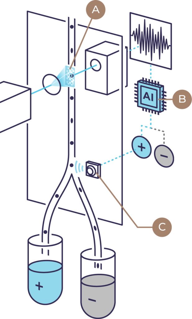
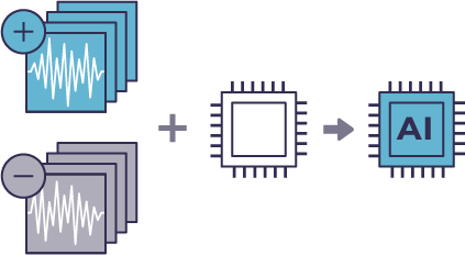
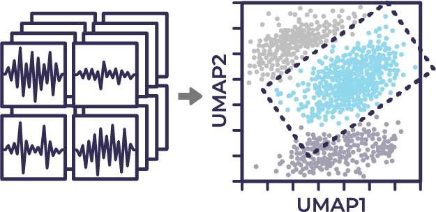
Science
Machine Vision-based
Cell Sorting
Characterizing and sorting cells based on image information at record high-throughput rates by integrating a novel ultrafast imaging technique with artificial intelligence.
How It Works
A) Image acquisition
Image information of each cell is recorded as a compressed temporal waveform with a single pixel detector as the cell passes through an illumination pattern projected onto a microchannel.

B) Machine
learning-based
classification
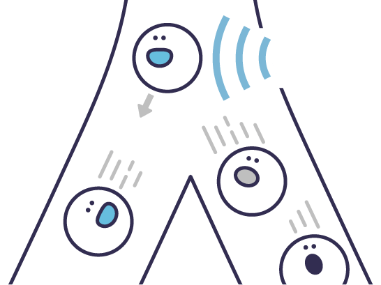
A trained AI model predicts the cell class based on the waveform.
C) Sorting
The classified cells are gently isolated using fluid pressure.
* Ota et al, Science, 2018 Jun 15;360(6394):1246-1251. doi: 10.1126/science.aan0096.
Core technology: Ghost Cytometry (GC) – Machine Learning-based Sorting

2D image information is collected as cells pass through a structured illumination via proprietary optic designs

… and recorded as a compressed 1D temporal waveform “Ghost Motion Imaging” (GMI)

GMI waveforms are directly analyzed by machine learning-based models for high-speed analysis and sorting, bypassing image reconstruction (first for classifier training then actual analysis of samples)

Target cells (both live and fixed) are gently collected using fluid pressure
Workflow of supervised
machine learning
The workflow outlines key steps involved in developing a machine learning model in ViCS technology.
1Labeling
Image information of each cell is acquired as a compressed temporal waveform using the novel imaging technique. The acquired waveforms are then labeled using biomarkers or other molecular labels.
2Modeling
A machine-learning model is developed based on the labeled waveforms.
3In Silico Labeling
The developed machine-learning model predicts labels by evaluating the waveforms of cells. This machine-learning approach enables image inference at an unprecedented speed.
4Profiling & Validation
Using the isolated cells, perform downstream analysis such as multi-omics analyses and functional assays to demonstrate applications.
Workflow of unsupervised
machine learning
1Image Data Acquisition
Image information of each cell is acquired as a compressed temporal waveform (GMI signals) using using Ghost Cytometry.
2Dimensional Reduction & Gating
Transform collected GMI signals into two-dimensional space using dimensional reduction methods such as t-SNE and UMAP and create gates to identify potentially unique subpopulations.
3Model Generation & Sorting
Create a classification model for the identified subpopulations and isolate them for downstream analysis such as single cell transcriptomic sequencing.
4Profiling & Validation
Using the isolated cells, perform downstream analysis such as multi-omics analyses and functional assays to demonstrate applications.