VCCT’s Challenge: Evaluating Cell Quality for Clinical Translation
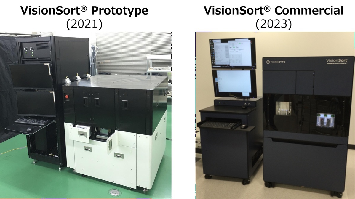
Guided by its vision, “Providing all solutions for all patients,” VC Cell Therapy Inc. (VCCT) is working to realize cell therapies for outer retinal diseases. At VCCT, Dr. Tomohiro Masuda leads manufacturing development and quality control, two functions central to the company’s R&D. To evaluate cell quality beyond conventional methods, Dr. Masuda introduced VisionSort®, a cell sorter equipped with ThinkCyte’s Ghost Cytometry® (hereafter GC) technology.
For cellular products, evaluating and managing cell characteristics is essential when defining release criteria. For teams working from basic research through clinical-grade products, this remains a persistent R&D challenge.
Even as two iPS-derived cellular products received their first conditional and time-limited approvals in Japan in 2026, methods for evaluating and managing cell properties remain at an early stage. For VCCT, one important question was whether cells could be evaluated non-invasively and in real time, without relying on viability stains or antibody-recognized surface antigens. An indicator like this could support R&D in cellular quality control, contribute to more stable manufacturing of effective cellular products, and help in the development and adoption of cell therapies.
This article describes how VCCT has used VisionSort to address practical research challenges in regenerative medicine. It covers the background to adoption, on-the-ground experience, observed value, and VCCT’s future outlook.
Index
- Challenges: Limits and Biases in Conventional Evaluation Methods
- The Gap Between Being ‘Alive’ and Being ‘Functional’
- The Bias of Predefined Markers in Antibody-Based Analysis
- Solutions: Label-Free and Morphological Analysis via GC
- A Novel Cell Sorting Method Powered by Morphological Information
- New Discoveries–Using Unsupervised Learning to Reveal New Patterns
- The Adoption Process: Co-development with ThinkCyte
-
- A Two-Stage Training Program
- Close Technical Support Throughout the Project
- Results: Establishing a New Evaluation Axis and Validation for Practical Use
-
- The Impact of Seeing Previously Hidden Differences
- Morphological Analysis as an Additional Evaluation Axis
- Future Outlook: Turning Confidence into Specifications
-
- Working Toward QC Specifications Based on Morphology
- Exploring the Molecular Basis Linking Morphology and Genetics
- Next Step–Who May Benefit from This Approach
Challenges: Limits and Biases in Conventional Evaluation Methods
For the VCCT research team, a key bottleneck was that QC evaluation of iPS-derived differentiated cells—their therapeutic tool—was largely limited to viability checks.
The Gap Between Being ‘Alive’ and Being ‘Functional’
“Conventional QC has primarily focused on measuring cell viability. However, in clinical practice, a more meaningful assessment is required: how healthy the living cells are, and how well they can function after transplantation. To help ensure the quality of transplanted tissue, our team has discussed the shared understanding that a new assay is needed—one that goes beyond conventional live/dead assessments and evaluates the qualitative value of the cells,” said Dr. Masuda.
The Bias of Predefined Markers in Antibody-Based Analysis
The team also evaluated antibody-based quality assessments using apoptosis, a form of cell death, as an indicator, but this approach had limitations. In conventional flow cytometry, antibody-based analysis requires researchers to define the target molecule in advance. For processes involving complex differentiation pathways or unknown phenotypes, relying only on predefined markers can limit the scope of analysis and what researchers are able to detect.
“To evaluate quality with less bias, we needed a different kind of technology,” said Dr. Masuda.
This need, born from scientific integrity, ultimately led to their encounter with GC technology.
Solutions: Label-Free and Morphological Analysis via GC
VCCT saw a possible way forward in ThinkCyte’s GC technology. The team first noticed the technology through a paper published in Science.
A Novel Cell Sorting Method Powered by Morphological Information
A defining feature of GC is that it uses AI to analyze cell morphology in real time without relying on conventional fluorescence data. Based on those morphological features, the system can sort target cells at high speed without staining, enabling label-free sorting.
For regenerative medicine applications such as VCCT’s retinal organoids, label-free sorting of target cells from heterogeneous populations can help support the manufacture of higher-purity therapeutic products while reducing physical and chemical stress on cells.
New Discoveries–Using Unsupervised Learning to Reveal New Patterns
For Dr. Masuda and the VCCT team, the deciding factor in adopting VisionSort, was its built-in AI analysis tools, including unsupervised learning-based visualization.
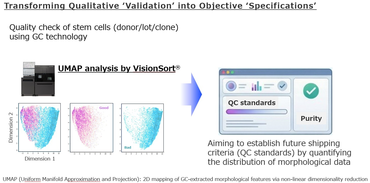
Unlike conventional flow cytometry, which categorizes cells based on fluorescence intensity of predefined markers, VisionSort can use UMAP to visualize subtle, difficult-to-define changes in cellular state as intuitive maps. For VCCT, this provided a way to extract objective data from cellular changes whose exact nature was unclear, but that were still apparent at the population level.
“Even when cell populations appear to show no clear difference at first glance, measuring them with VisionSort and applying UMAP analysis can reveal clear shifts in population distribution or the appearance of distinct ‘islands’. This suggests that morphological information may reveal cell-state changes in specific subpopulations that conventional indicators could not fully capture,” said Dr. Masuda.
In early-stage research and marker discovery, the ability to analyze morphological information in an unbiased way—rather than relying only on predefined antibody markers—is expected to offer a significant advantage.
The Adoption Process: Co-development with ThinkCyte
Although introducing and establishing a technically novel analytical instrument in a research environment can present challenges, the VisionSort implementation process was carried out step by step, with comprehensive support throughout.
A Two-Stage Training Program
Before installing VisionSort at VCCT, the team had the opportunity to use the instrument at ThinkCyte’s facility on the University of Tokyo campus (back then) until they were fully comfortable with it.
“We followed a two-stage process: first, we learned the basics using a demonstration unit at ThinkCyte’s facility. Then, after the instrument was installed at our own facility, we continued training with our own cells. This seamless transition from basic operation to hands-on use with helped build proficiency more quickly and enabled smooth implementation into our workflow,” said Dr. Masuda.
Close Technical Support Throughout the Project
“In addition to hardware installation, ThinkCyte’s engineering and research teams provided detailed reports after experiments and held careful planning meetings in advance. This close support helped us move the research project forward,” said Dr. Masuda.
For VCCT, this reflected not only support for implementing the instrument, but also a partnership aimed at joint technology development and real-world adoption.
Results: Establishing a New Evaluation Axis and Validation for Practical Use
Before full installation, VCCT and collaborators sorted retinal progenitor cells label-free using a prototype VisionSort device, and the results were published in a peer-reviewed paper. After VisionSort was formally introduced, it added a new perspective to VCCT’s research workflow.
The Impact of Seeing Previously Hidden Differences
In VCCT’s case, the moment when the team felt the greatest value in practice came from the UMAP analysis results.
“When sample-level differences were present only in a small subpopulation, supervised learning did not always produce a high discrimination score, such as ROC-AUC. However, when we analyzed the same data using UMAP, those differences became visible in the position of the clusters and the spread of their distributions,” said Dr. Masuda.
Morphological Analysis as an Additional Evaluation Axis
“When comparing cell phenotypes, considering the possibility of a morphological change and using VisionSort to check, is becoming an option alongside conventional analytical methods,” said Dr. Masuda.
In VCCT’s workflow, this could help the team routinely screen and rescue cellular states that might previously have been overlooked or left unevaluated
If a framework to evaluate quality differences based on morphological data can be established, it can support more accurate project decision-making and may contribute to lower manufacturing costs, improved reliability, and greater confidence in regulatory planning. Even in early-stage research where appropriate markers have not yet been established, VisionSort can help VCCT grasp the morphological landscape of a cell population. Furthermore, analyzing distinctive cell populations separated on a UMAP plot may provide a path toward defining future QC indicators.
“Our upcoming challenge is to establish the qualitative impact brought by VisionSort—the experience of ‘seeing what was once invisible’—as objective, quantitative metrics,” said Dr. Masuda.
This suggests that VisionSort may demonstrate its true value as a powerful exploratory tool for identifying unknown biomarkers and cellular states. At research stages where appropriate markers have not yet been established, it can provide an unbiased view of the overall cell population and offer a path toward extracting new features that may serve as future quality control indicators. The previously unseen cellular profiles revealed by this exploratory tool are becoming an essential starting point for the next challenges of quantification and standardization.
Future Outlook: Turning Confidence into Specifications
VCCT’s work with VisionSort is still at an early stage. Dr. Masuda has set “moving from qualitative to quantitative” as a milestone for the next one to two years.
Working Toward QC Specifications Based on Morphology
VCCT is currently at the stage of gaining qualitative confidence that “differences are visible”; the next hurdle is converting those differences into objective numerical values that could later inform release criteria or manufacturing process control indicators. This would involve quantifying distribution shifts or specific cluster ratios on a UMAP plot and using accumulated data to examine how those values correlate with post-transplantation engraftment rates and functional retention.
“We intend to eventually quantify the qualitative differences shown by VisionSort into objective numerical values, polishing them into rigorous QC specifications for our manufacturing process,” said Dr. Masuda.
Exploring the Molecular Basis Linking Morphology and Genetics
VCCT also plans to use VisionSort’s high-speed sorting capabilities to isolate morphologically distinct cell populations and then analyze them with single-cell gene expression and other tests.
If the biological meaning underlying AI-captured morphological differences can be decoded, GC could develop into a non-invasive quality-assessment tool for regenerative medicine.
Next Step–Who May Benefit from This Approach
Based on his experience, Dr. Masuda notes that VisionSort may help researchers facing the following challenges:
- Handling heterogeneous cell populations.
- Lacking surface markers or facing the limitations of antibody-based analysis.
- In the early exploratory stages of a project and wanting to first gain an overall view as a starting point.
- Seeking non-invasive cell-evaluation indicators from genetic cell-marking data, such as GFP-based genetic markers generated with CRISPR-Cas9 or similar tools.
“I feel that one effective approach would be to first use VisionSort to gain an overall view, and then look for features that could serve as markers,” said Dr. Masuda.
- Want to learn more? Click here for other additional iPS cell analysis case studies.
- Want to understand technology? Click here to explore publications on GC technology and its applications.
- Want to test your own samples? Consult with us here to discuss how GC could address your research challenges.
VCCT’s experience suggests that a new instrument can do more than replace an existing tool. In this case, VisionSort added another dimension of insight to the team’s research.
- Routinely incorporating morphological information into cell QC may help researchers make more informed, quality-based selections of cellular products.
- Capturing the overall picture of cell populations before markers are established may support the exploration of unknown biomarkers and cellular states.
In regenerative medicine, evaluating cell quality is closely connected to the safety and trust required for therapies delivered to patients. For VCCT, VisionSort offers a way to support work from exploratory research through manufacturing development.